Quick Contact
+91 97956 17171
+022 35631299
reemshadiagnostics@gmail.com
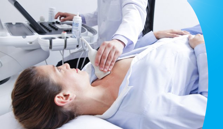
Sonography
Ultrasound scans use sound waves to build up a picture of the inside of the body. They are completely painless. The ultrasound scanner has a probe that gives off sound waves. The probe is passed over your body. The sound waves bounce off the organs inside your body and are picked up again by the probe. The probe is linked to a computer. This turns the reflected sound waves into a picture.
“Ultrasound” merely means sound waves with a frequency higher than 20 kHz, which is the highest range that the average human can hear. Modern sonography equipment utilizes these high-frequency sound waves to create high-resolution images of the inside of the human body from soft tissues to organs.
Ultrasonography
Ultrasound imaging uses sound waves to produce pictures of the inside of the body. It is used to help diagnose the causes of pain, swelling, and infection in the body’s internal organs and to examine a baby in pregnant women and the brain and hips in infants. It is also used to help determine and perform biopsies, heart conditions, and damage after a heart attack.
Ultrasonography is commonly used to evaluate the following:
Abdomen: Abdominal ultrasound can help to detect problems in the : blood Vessels in the abdomen Gallbladder Intestines Kidneys Liver Pancreas Spleen
An Abdominal ultrasound is commonly used to evaluat the couse of stomach pain, bloating, to check for kidney stones, liver diseases, tumors and many other conditions.
Blood vessels: For example, to detect dilated and narrowed blood vessels.
Urinary tract: For example, to distinguish benign cysts from solid masses in the kidneys or to detect blockages such as stones or other structural abnormalities in the kidneys, ureters, or bladder.
More information
Your doctor will usually tell you to fast for 8 to 12 hours before your ultrasound. That's because undigested food in the stomach and urine in the bladder can block the sound waves, making it difficult for the technician to get a clear picture.
Ultrasonography uses high-frequency sound (ultrasound) waves to produce images of internal organs and other tissues. A device called a transducer converts electrical current into sound waves, which are sent into the body's tissues.
Types of Ultrasound:
Endoscopic ultrasound.
Doppler ultrasound.
Color Doppler.
Triplex ultrasound (color-flow imaging)
Transvaginal ultrasound.
Duplex ultrasound.
Reemsha Diagnostics center is one of the most trusted Diagnostic centers in Navi Mumbai with over 05 years of expertise, excellence, and empathy in health care services.
Contact Us

+91 97956 17171
+022 35631299

reemshadiagnostics@gmail.com

Shop no. 5 and 6, Patel Heights,Plot No. 15, 16 and 17, Opp. Rajeev Gandhi College, Sector 7, Ghansoli, Navi Mumbai, Maharashtra 400701.